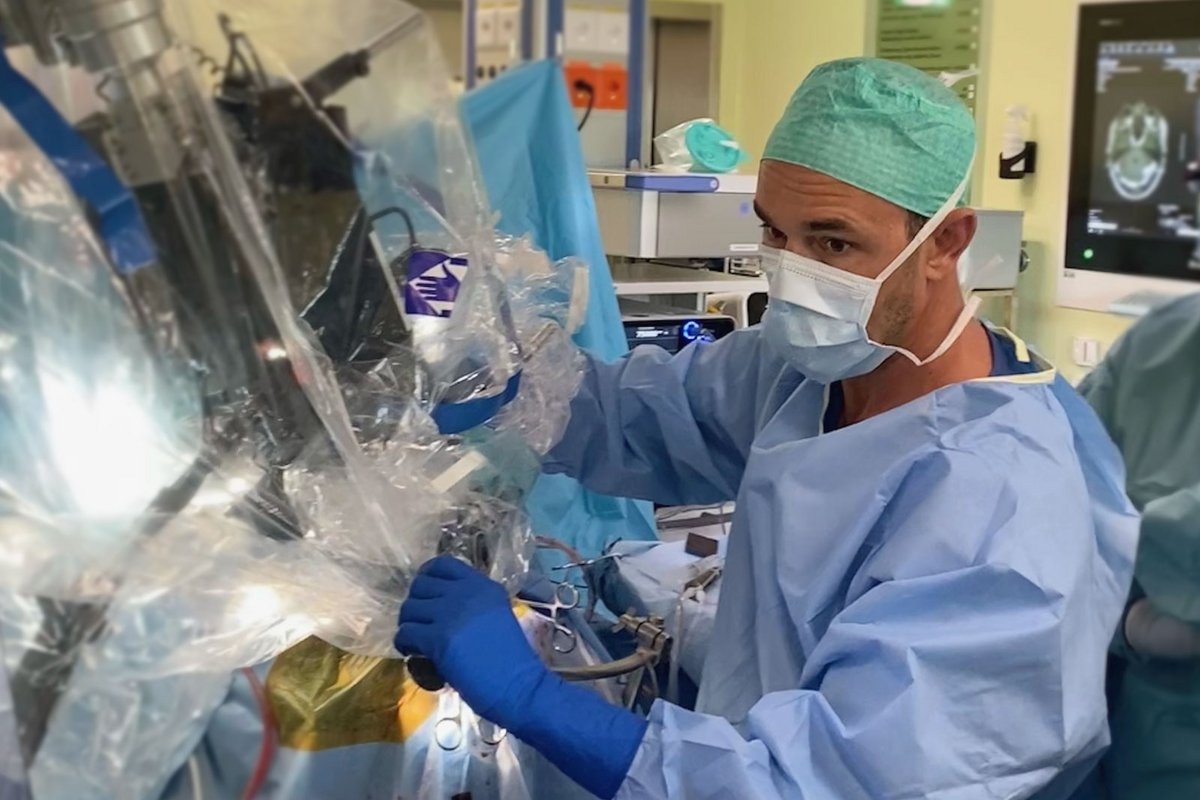
Another major milestone of the HORAO research project has been reached: a newly developed type of surgical microscope is being used for the first time during brain tumor surgery at the Inselspital Bern. With the help of innovative imaging, it should enable a more radical and at the same time safer removal of brain tumors. Initial measurements show promising results.
What is HORAO about?
After a brain tumor has been diagnosed, complete resection is the first and crucial step in the treatment plan. However, even with the most modern surgical microscopes, it is still difficult to distinguish healthy brain tissue from diseased tumor tissue during surgery and to identify important fiber tracts that carry out vital functions. There is a high risk that a tumor cannot be completely removed or that neurological damage will occur after surgery.
This is precisely where the HORAO research project comes in. Specialists in optical physics, pathology, neurosurgery and artificial intelligence are working together to develop a new optical method that will lead to safer, faster and more precise tumor resections.
The technology behind it
HORAO uses the technology of Mueller polarimetry, or IMP for short. This is based on the fact that highly organized structures in the brain reflect polarized light differently than the unorganized cells of tumors. Mueller polarimetry also has the advantage that it can be applied in real time, non-invasively and without the use of contrast agents. However, in order for this to be used in the operating room, the IMP must be miniaturized to such an extent that it can be built into a neurosurgical operating microscope at the end of the development process. With the help of state-of-the-art computer visualization, automated tumor segmentation should then be possible. The fiber tracts, which are the carriers of important brain functions, can also be visualized under polarized light and spared during surgery.
Crowdfunding and crowdsourcing – ideas for HORAO
HORAO was born out of the minds of a few creative neurosurgeons at the Insel Hospital. They were looking for an answer to the question: How can healthy brain tissue be safely distinguished from tumor tissue? A global brainstorming, a so-called crowdsourcing competition, was designed to bring them closer to solving this medical problem. After the first Swiss crowdfunding in the field of medical research to generate funds, the crowdsourcing was completed in 2019 and the HORAO project by Ivan Gusachenko from France was chosen as the winner. Two years later, the international research team around HORAO received the prestigious Sinergia Grant from the SNSF, worth 2.3 million Swiss francs, which fully funds the research project for 4 years.
Who is behind HORAO?
At HORAO, two international research teams from four institutions are working closely together. They include scientists from the Ecole polytechnique in Paris, Swiss doctors specializing in neurosurgery and neuropathology, experts in artificial intelligence and machine learning, and optical engineers. Prof. Philippe Schucht, MD, PhD, deputy head of the neurosurgery department and head of neuro-oncology at Inselspital in Bern, is the project manager. David Hasler is responsible for project coordination.
What are the next steps?
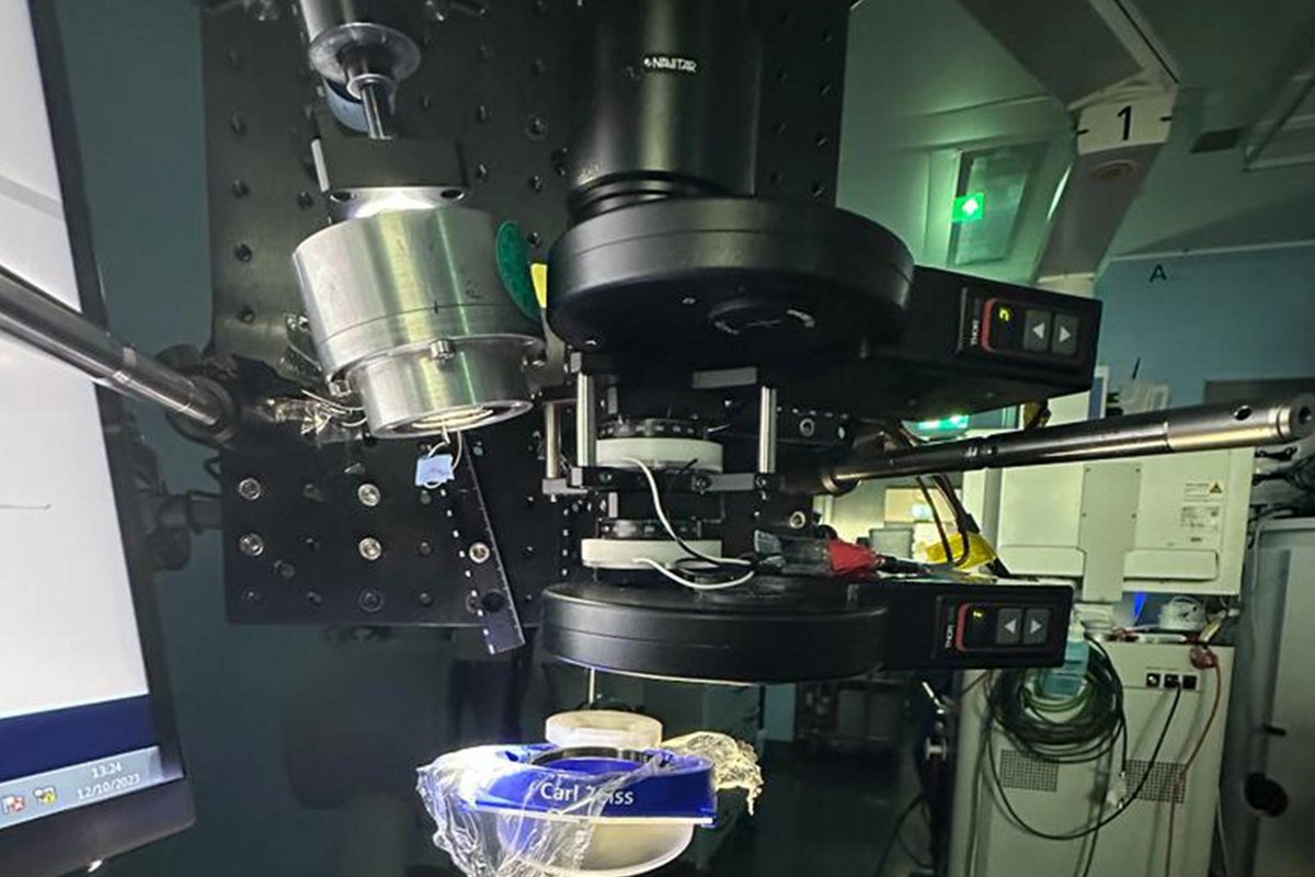
With the first test applications in the operating room, an important milestone has been reached. However, there is still a long way to go for the researchers at HORAO. The current model is a simple prototype that needs to be further miniaturized to achieve a small final size of the device. The measurement accuracy needs to be improved, as do real-time measurements, which are currently not possible. The hardware and software are being patented in parallel.
The research team is currently in discussions with several potential industrial partners who are to be brought on board for the development of an industry-ready microscope model. The completion of HORAO as a research project is currently planned for 2026.
Related News
- SNF-Grant in Millionenhöhe für Prof. Philippe Schucht und sein HORAO-Projekt23.12.21 - Grosse Auszeichnung für Prof. Dr. med. Philippe Schucht, Stv. Chefarzt Neurochirurgie und Leiter Neuroonkologie, und sein Team.
- Ivan Gusachenko gewinnt HORAO15.03.19 - An der HORAO-Konferenz vom 14. März 2019 haben die Jury und das Publikum das Siegerprojekt gekürt. Ivan Gusachenko aus Frankreich hat den globalen…
- HORAO erhält 45 Projekteingaben19.11.18 - Die Universitätsklinik für Neurochirurgie hat für ihr prämiertes Projekt HORAO im globalen Crowdsourcing-Wettbewerb 45 Projekteingaben aus 23…
- Erfolgreiches Crowdfunding19.09.17 - Die Universitätsklinik für Neurochirurgie am Inselspital Bern hat es geschafft: Sie hat die Crowdfunding-Kampagne zum Projekt HORAO erfolgreich…