
Neuronavigation is a computer-assisted procedure that enables precise navigation in the brain and spine. Images from computed tomography or magnetic resonance imaging are processed using special software to create three-dimensional models that are used during surgery. This allows anatomical structures and instrument guidance to be displayed directly on the screen—even during complex spinal procedures.
How has spinal neuronavigation evolved?
Spinal neuronavigation developed from frameless stereotaxy on the skull. Since the spine is very mobile, precise navigation was difficult at first. From the mid-1990s onwards, anatomical landmarks directly on the spine significantly improved accuracy *, *. CT and X-ray-based procedures were later added, in which image data is transferred directly to the navigation system. These advances now enable high-precision procedures on the entire spine.
Improved precision
Spinal neuronavigation increases safety and accuracy in spinal surgery. Pedal screws are used in many spinal surgeries. These are special screws that are inserted into the vertebrae to stabilize the spine.
Studies show that screws can be placed much more precisely with the aid of navigation. In a large-scale analysis, the risk of misplacement was only 6%, whereas it was 15% with the conventional method—with no neurological complications *.
Navigation-assisted placement of pedicle screws is more accurate than conventional placement under fluoroscopy or using anatomical landmarks («freehand technique»). With navigation, pedicle screws can be positioned clinically correctly in 96–99% of cases, compared to 93–94% with conventional methods.
The most conclusive scientific data come from a meta-analysis conducted in 2024. This analysis evaluated the results of 30 studies involving a total of 17,911 patients and 24,600 pedicle screws. The analysis showed that navigation-guided screw placement achieved a clinically correct screw position in 96.2% of cases, compared with 94.2% for conventional methods. This difference was statistically significant. *
Other studies also confirm that navigation enables more reliable and safer screw placement *, *.
Lower radiation exposure
An additional advantage is the significantly lower radiation exposure: thanks to the navigation system, the radiation dose for both patients and the surgical team can be reduced to a minimum *.
How is spinal neuronavigation used?
There are different spinal neuronavigation systems.
In the past, preoperative computed tomography images were combined with 3D X-ray images taken during surgery.
Today, modern navigation software can directly compare the surface of the vertebral body with previously created CT data and transfer the data to the operating room monitor during surgery. This allows the surgeon to see the position of the instruments and of the screws in real time in a three-dimensional image of the spine. This makes spinal surgery even more precise and safer.
At Inselspital: O-arm
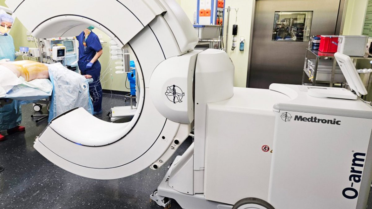
For instrumented spinal surgery, we routinely use the O-arm. This is a mobile X-ray system that generates high-resolution two- and three-dimensional images of the spine during surgery. These images can be combined with a navigation system. This allows the surgeon to place instruments and implants—such as pedicle screws—with exceptional precision in real time.
Advantages of the O-arm Navigation System
- High-resolution 3D imaging directly during surgery
- High precision in the placement of pedicle screws
- Immediate verification of implant position with the ability to correct it directly if necessary
- Fewer postoperative follow-up CT scans required
- Particularly helpful for complex deformities, revision surgeries, or significantly altered anatomy
-
Shin BJ, James AR, Njoku IU, Härtl R. Pedicle screw navigation: a systematic review and meta-analysis of perforation risk for computer-navigated versus freehand insertion. J Neurosurg Spine. 2012;17:113-122.
-
Richter M, Cakir B, Schmidt R. Cervical pedicle screws: conventional versus computer-assisted placement of cannulated screws. Spine (Phila Pa 1976). 2005;30:2280-2287.
-
Papalia GF, Vadalà G, Russo F, Marcello G, Nardi N, Papalia R, Denaro V. Higher Accuracy and Better Clinical Outcomes in Navigated Thoraco-Lumbar Pedicle Screw Fixation Versus Conventional Techniques : A Systematic Review and Meta-Analysis. Spine (Phila Pa 1976). 2024 Oct 1;49(19):1370-1380. doi: 10.1097/BRS.0000000000005105. Epub 2024 Jul 25.
-
Fichtner J, Hofmann N, Rienmüller A et al. Revision Rate of Misplaced Pedicle Screws of the Thoracolumbar Spine-Comparison of Three-Dimensional Fluoroscopy Navigation with Freehand Placement: A Systematic Analysis and Review of the Literature. World Neurosurg. 2018;109:e24-e32.
-
Schuetze K, Eickhoff A, Dehner C, Schultheiss M, Gebhard F, Richter PH. Radiation exposure for the surgical team in a hybrid-operating room. J Robot Surg. 2019;13:91-98.