
Pituitary adenomas are benign tumors of the pituitary gland, which is located at the base of the skull. A distinction is made between hormone-secreting and non-hormone-secreting adenomas, as well as microadenomas (smaller than 1 cm) and macroadenomas (1 cm or larger). Pituitary adenomas can cause hormonal disorders and visual disturbances. For prolactinomas, medication is the treatment of choice. For all other pituitary adenomas, surgical removal through the nose is generally indicated. This minimally invasive procedure is performed at Inselspital using endoscopic techniques.
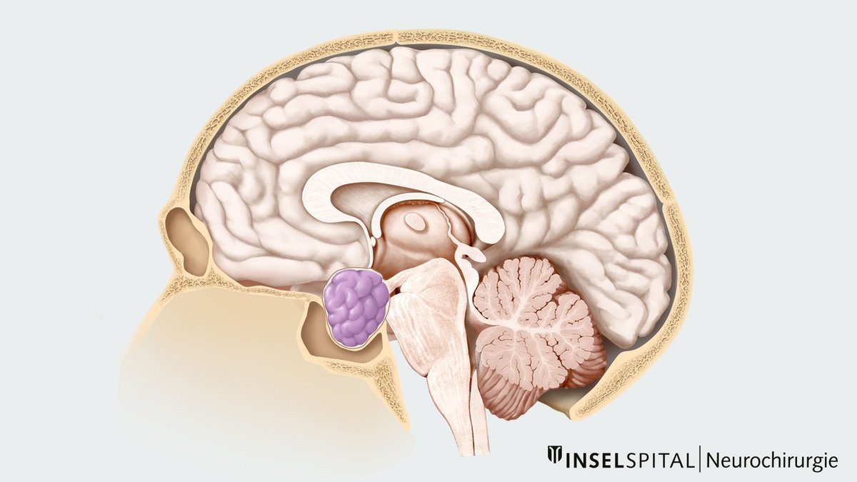
How common is a pituitary adenoma?
Pituitary adenomas account for about 15% of all brain tumors * and are thus among the most common tumors of the nervous system. In about one in 1,100 people, the lead to medically significant symptoms or findings. Women are affected slightly more often than men *.
Pituitary adenomas can occur at any age. Their incidence increases slightly with age and is approximately 8 cases per 100,000 people per year among those over 65. Very small pituitary adenomas, known as microadenomas, are often discovered by chance. They are found in about 10–15% of the population *, * and in many cases have no impact on health. In such cases, regular follow-up is usually sufficient.
What is the difference between a macroadenoma and a microadenoma?
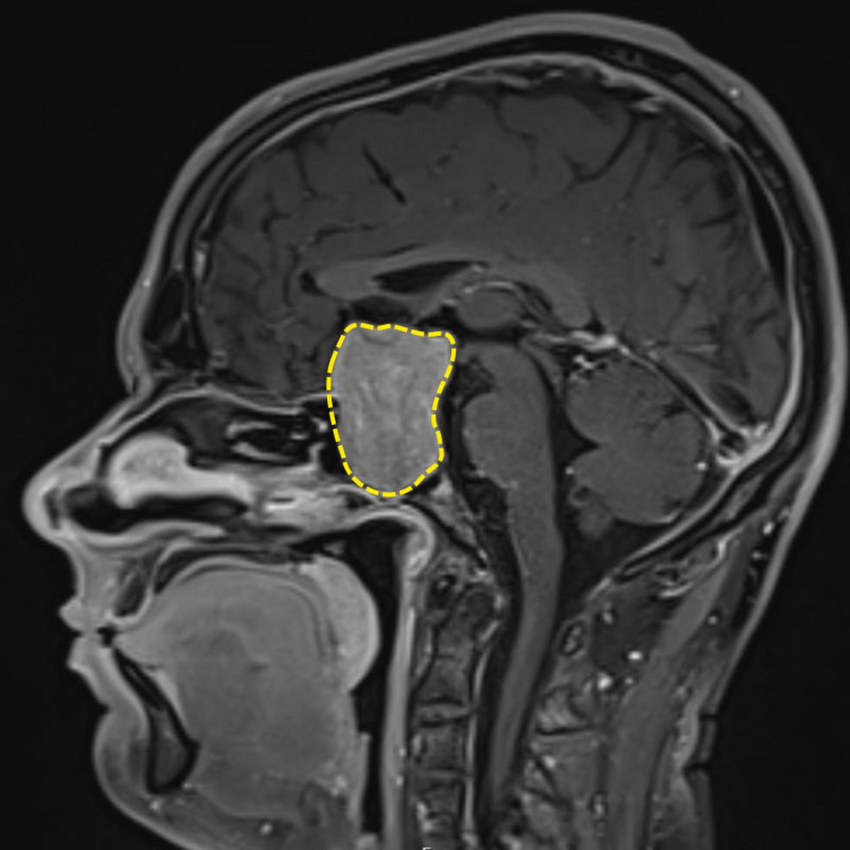
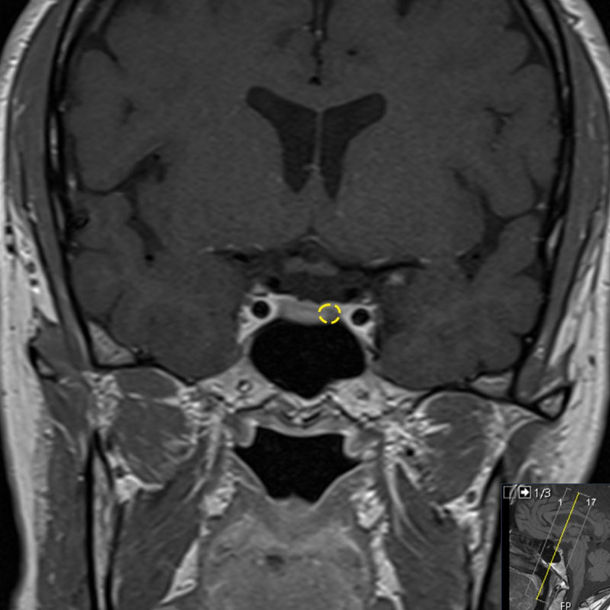
The main difference lies in the tumor size: macroadenomas are at least 1 cm in size, while microadenomas are smaller than 1 cm. About half of the pituitary adenomas that cause symptoms or other medically significant abnormalities are macroadenomas.
This distinction is important for two main reasons:
- Compression of adjacent structures: Due to their size, macroadenomas can exert pressure on adjacent structures. The optic nerves, in particular, can be affected.
- Tumor growth: Macroadenomas are more likely to continue growing. The probability of further growth is approximately 25% within 4–5 years *, *.
What symptoms can a pituitary adenoma cause?
Small, non-hormone-secreting pituitary adenomas usually do not cause any symptoms. They are often discovered incidentally when imaging of the head is performed for another reason.
Larger or hormone-secreting pituitary adenomas can cause various symptoms, depending on their size and hormone production:
- Hormonal disorders: overproduction or deficiency of certain hormones
- Pressure on adjacent structures: for example, causing visual disturbances or impairment of cranial nerves
- Pituitary apoplexy: a sudden hemorrhage into the pituitary adenoma
Hormonal disorders
Hormonal disorders are caused either by excessive hormone production due to a pituitary adenoma or by insufficient hormone production due to a dysfunction of the healthy pituitary gland.
| 30% | do not produce hormones (non-hormone-secreting adenomas) |
| 53% | produce the hormone prolactin (prolactinoma) |
| 12% | produce growth hormone and lead to acromegaly |
| 4% | produce the hormone ACTH and lead to Cushing's disease |
| 1% | produce thyrotropin (TSH), luteinizing hormone (LH), or follicle-stimulating hormone (FSH) |
Hormone-producing tumors can cause different clinical presentations depending on the hormone they produce. For more information on the possible symptoms, please refer to the descriptions of the respective clinical presentations.
Depending on the size of the tumor, pituitary dysfunction occurs in about half of patients. This can lead to a deficiency of various pituitary hormones:
- Deficiency of LH and FSH (hypogonadism): As a result, the body produces too few sex hormones. In women, this can lead to irregular or absent menstruation. In both women and men, symptoms may include fatigue, lack of motivation, loss of libido, and infertility.
- Deficiency of TSH (hypothyroidism): This causes the thyroid gland to produce too few thyroid hormones. Typical symptoms include increased sensitivity to cold, weight gain, fatigue, digestive problems, skin changes, and memory problems.
- ACTH deficiency (hypercortisolism): This causes the adrenal cortex to produce too little cortisol. Possible symptoms include low blood pressure—especially upon standing—severe fatigue, nausea and vomiting, dizziness, and abdominal pain. A severe cortisol deficiency can lead to a life-threatening adrenal crisis (Addisonian crisis). Cortisol is a vital hormone and plays a central role in the body’s adaptation to stressful situations. A cortisol deficiency must be treated urgently with medication.
Pressure on adjacent brain structures
A macroadenoma can press on adjacent brain structures and nerves, thereby causing symptoms. In addition to headaches, these include:
- Visual disturbances: Visual acuity may decrease, and the visual field may be restricted. If the adenoma presses on the optic chiasm, the outer areas of the visual field in both eyes are typically lost. This visual disturbance is known as bitemporal hemianopia.
- Cranial nerve impairment: If the adenoma invades the adjacent cavernous sinus, double vision, facial pain, or drooping of the upper eyelids (ptosis) may occur.
- Impaired cerebrospinal fluid drainage: In rare cases, the adenoma can block the drainage of cerebrospinal fluid. This results in what is known as obstructive hydrocephalus, in which intracranial pressure rises. Possible signs include headaches, vomiting, and altered consciousness.
How is a pituitary adenoma diagnosed?
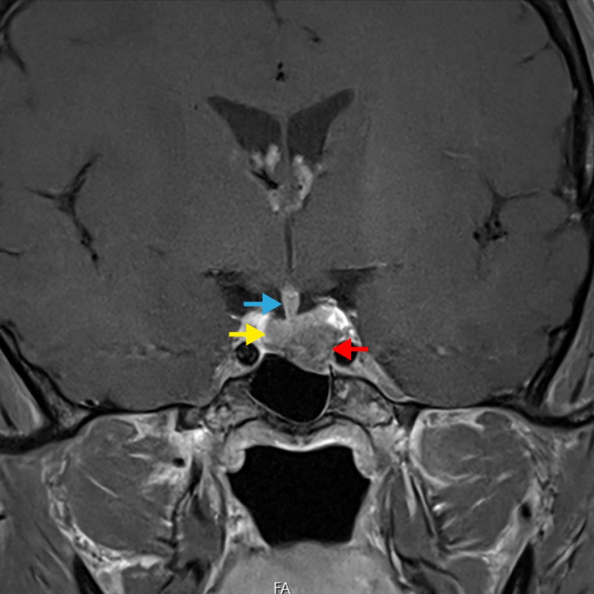
Magnetic resonance imaging (MRI) with contrast medium is the imaging method of choice. A special pituitary protocol is used to ensure an accurate evaluation.
To reliably detect even very small tumors, the examination may be supplemented with a dynamic MRI sequence in certain cases. In this procedure, cross-sectional images of the pituitary gland are acquired at several time points (for example, at 0, 30, 60, and 90 seconds). Small pituitary adenomas often take up the contrast agent more slowly than healthy pituitary tissue, making them easier to detect. Dynamic MRI sequences can also be helpful after surgery to distinguish small tumor remnants from scar tissue. Particularly in the case of small adenomas, such as those that can occur in Cushing’s disease, these specialized sequences can significantly increase the likelihood of detection *, *.
Hormone levels are also tested in all patients with a pituitary adenoma. This allows for the identification of potential hormonal disorders and their treatment if necessary. This evaluation is typically performed by a specialist in endocrinology.
If imaging shows that the adenoma is affecting the optic nerves or their crossing point (optic chiasm), an additional ophthalmological examination is required. During this examination, visual acuity and the visual field are assessed. If the adenoma is located near the optic tracts, the examination also serves to establish a baseline for future comparisons.
For treatment planning, it is also important to determine whether and to what extent the adenoma has invaded the adjacent venous blood vessel, the cavernous sinus. The extent of this invasion is assessed using the so-called Knosp classification.
Petrosal sinus sampling
In rare cases, if Cushing’s disease is suspected, an additional invasive test may be necessary: a procedure known as petrosal sinus sampling. It is used in particular when the results of hormone tests and MRI scans do not provide a clear picture.
In this procedure, thin catheters are inserted through a vein in the groin and advanced into the venous sinuses of the pituitary gland. There, the concentration of the hormone ACTH is measured and compared to the concentration in the rest of the bloodstream. An ACTH-producing pituitary adenoma releases the hormone directly into these venous sinuses.
If the ACTH concentration measured there is at least twice as high as in the rest of the bloodstream, this strongly suggests that the pituitary gland is the source of the elevated ACTH production. After administration of the stimulating hormone CRH, a value at least three times higher is considered a corresponding indication. The result thus supports the diagnosis of Cushing’s disease *, *.
The test may also provide clues as to which side of the pituitary gland the adenoma is located on. However, this determination is not always reliable. The venous drainage system of the pituitary gland can vary from person to person and may therefore influence the test result *, *, *.
Functional imaging: amino acid PET
In selected cases, an amino acid PET scan may be performed as a supplementary test for hormone-producing pituitary adenomas. This examination is conducted at the Department of Nuclear Medicine. During the procedure, a weakly radioactively labeled amino acid – known as a tracer – is administered intravenously. This tracer accumulates preferentially in the tumor tissue. A PET scanner detects the signals emitted by the tracer. The PET images are then combined with the MRI images. This allows for better visualization of metabolically active tumor tissue *, *, *.
An amino acid PET scan can be particularly helpful if
- the tumor cannot be clearly identified or delineated on MRI,
- the results of hormone tests and the MRI do not match, or
- disease activity persists after surgery.
However, because the spatial resolution is lower than that of an MRI, the test has its limitations. Its use in the diagnosis of pituitary adenomas is still being scientifically investigated and is not yet part of routine diagnostic practice.
Computed tomography of the skull
A computed tomography (CT) scan of the skull provides important information for surgical planning. We routinely perform this scan prior to pituitary surgery.
The scan reveals the individual bony structures and any possible anatomical variations. This allows us to precisely plan the surgical approach and optimally prepare for the operation.
When is genetic testing recommended?
Genetic testing is necessary only in exceptional cases. The test specifically looks for a germline mutation in the MEN1 gene. Genetic testing should be considered, in particular, in the following situations:
- in individuals aged 30 or younger with a hormone-secreting pituitary adenoma (except for women with a prolactinoma)
- in individuals aged 30 or younger with a macroadenoma (at least 1 cm)
- in children and adolescents aged 18 or younger with a pituitary adenoma
- in cases of MEN-associated tumors in the affected individual or in first-degree relatives
How are pituitary adenomas treated?
Treatment depends on various factors. The appropriate therapy for each condition is explained below.
Why you should seek treatment at Inselspital
- Interdisciplinary team: The Neurosurgery Department at Inselspital works closely with the departments of Endocrinology, Ophthalmology, Ear, Nose, and Throat Medicine and Radiation Oncology.
- Pituitary Board: Every case is discussed by our interdisciplinary Pituitary Board. Together, the specialists develop an individualized treatment plan for each patient.
- Modern Intraoperative Technologies: During surgery, we make targeted use of modern technical tools.
- Endoscopic Surgical Technique: We generally perform pituitary surgeries endoscopically, that is, using a camera. The departments of Neurosurgery and Otolaryngology (ENT) work closely together in this process.
- Extended surgical approaches: Our surgical experience and close interdisciplinary collaboration enable us to perform even complex, extended procedures. These include, for example, the removal of the wall of the adjacent venous sinus (medial wall of the cavernous sinus) that faces the tumor. Particularly in cases of acromegaly and Cushing’s disease, this procedure can increase the chances of complete tumor removal and cure.
Resection of the medial wall of the cavernous sinus - Preservation of nasal function: The condition of the nose has a significant impact on quality of life. Approximately 10,000 liters of air flow through the nose every day. For this reason, we place great emphasis on preserving the structures and function of the nose and ensuring careful nasal care following surgery.
-
Ostrom QT, Gittleman H, Xu J, Kromer C, Wolinsky Y, Kruchko C, et al. CBTRUS Statistical Report: Primary Brain and Other Central Nervous System Tumors Diagnosed in the United States in 2009-2013. Neuro Oncol. 2016;18(suppl_5):v1-v75.
-
Tritos NA, Miller KK. Diagnosis and Management of Pituitary Adenomas: A Review. Jama. 2023;329(16):1386-98.
-
Ezzat S, Asa SL, Couldwell WT, Barr CE, Dodge WE, Vance ML, et al. The prevalence of pituitary adenomas: a systematic review. Cancer. 2004;101(3):613-9.
-
Fernández-Balsells MM, Murad MH, Barwise A, Gallegos-Orozco JF, Paul A, Lane MA, et al. Natural history of nonfunctioning pituitary adenomas and incidentalomas: a systematic review and metaanalysis. J Clin Endocrinol Metab. 2011;96(4):905-12.
-
Huang W, Molitch ME. Management of nonfunctioning pituitary adenomas (NFAs): observation. Pituitary. 2018;21(2):162-7.
-
Bashari WA, Senanayake R, Fernández-Pombo A, Gillett D, Koulouri O, Powlson AS, et al. Modern imaging of pituitary adenomas. Best Pract Res Clin Endocrinol Metab. 2019;33(2):101278.
-
Grober Y, Grober H, Wintermark M, Jane JA, Oldfield EH. Comparison of MRI techniques for detecting microadenomas in Cushing's disease. J Neurosurg. 2018;128(4):1051-7.
-
Micko AS, Wöhrer A, Wolfsberger S, Knosp E. Invasion of the cavernous sinus space in pituitary adenomas: endoscopic verification and its correlation with an MRI-based classification. J Neurosurg. 2015;122(4):803-11.
-
Eulate-Beramendi S, Casajús A, Ollero L, Niemann LK, Fernández-Miranda JC, Bruneau M, et al. Update in Cushing disease: What the neurosurgeon has to KNOW, on behalf of the EANS skull base section. Brain Spine. 2022;2:100917.
-
Pecori Giraldi F, Cavallo LM, Tortora F, Pivonello R, Colao A, Cappabianca P, et al. The role of inferior petrosal sinus sampling in ACTH-dependent Cushing's syndrome: review and joint opinion statement by members of the Italian Society for Endocrinology, Italian Society for Neurosurgery, and Italian Society for Neuroradiology. Neurosurg Focus. 2015;38(2):E5.
-
Ardakani MT, Rabizadeh S, Yadegar A, Mohammadi F, Reyhan SK, Qahremani R, et al. Bilateral inferior petrosal sinus sampling: validity, diagnostic accuracy in lateralization of pituitary microadenoma, and treatment in eleven patients with Cushing's syndrome - a single-center retrospective cohort study. BMC Endocr Disord. 2023;23(1):232.
-
Martini ML, Ransom RC, Rechberger JS, O'Keeffe D, Young W, Atkinson JLD, et al. Angiographic cross-filling between inferior petrosal sinuses and alteration of adrenocorticotropic hormone sampling results for tumor localization in Cushing disease. J Neurosurg. 2024;140(2):386-92.
-
Webb KL, Hinkle ML, Walsh MT, Bancos I, Shinya Y, Van Gompel JJ. Inferior Petrosal Sinus Sampling Tumor Lateralization and the Surgical Treatment of Cushing Disease: A Meta-Analysis and Systematic Review. World Neurosurg. 2024;182:e712-e20.
-
Flaus A, Levigoureux E, Haesebaert J, Briet C, Castinetti F, Cristante J, et al. Prospective Multicenter Evaluation of [(11)C]Methionine PET/MRI Sensitivity Compared with MRI for Localizing Small Pituitary Neuroendocrine Tumor or Pituitary Adenoma in Cushing Disease. J Nucl Med. 2025;66(10):1575-80.
-
Furnica RM, Devuyst F, Mathey C, Constantinescu SM, De Herdt C, Alexopoulou O, et al. Diagnostic Value of 11C-Methionine PET-CT Imaging in Persistent or Recurrent Cushing Disease After Surgery. J Clin Endocrinol Metab. 2025;110(10):2845-52.
-
Pruis IJ, Verburg FA, Balvers RK, Harteveld AA, Feelders RA, Vernooij MW, et al. [(18)F]FET PET/MRI: An Accurate Technique for Detection of Small Functional Pituitary Tumors. J Nucl Med. 2024;65(5):688-92.