
A traumatic brain injury occurs when the brain is injured as a result of a fall, a blow, or a traffic accident. Most head injuries in children are minor and heal without any lasting effects. However, if warning signs such as loss of consciousness, repeated vomiting, increasing drowsiness, or severe headaches are present, prompt medical evaluation is necessary. At Inselspital, children with traumatic brain injury are examined, monitored, and treated as part of an interdisciplinary approach.
How common is traumatic brain injury?
Head injuries are among the most common reasons why children are brought to an emergency department *, *. The vast majority have a mild traumatic brain injury.
Even among children who undergo more detailed evaluation, including a computed tomography (CT) scan, only about 6 to 8% are found to have an injury inside the skull. The findings are clinically significant in less than 2% *, *. Surgery is required in less than 1% of the children examined by CT *. Bleeding is therefore more of an exception, and most detected bleeds do not require surgery.
What are the causes of traumatic brain injury?
A traumatic brain injury is caused by external force applied to the head, such as:
- a fall
- a blow to the head
- a sports or recreational accident
- a traffic accident
In addition to brain injury, this can result in a skull fracture or bleeding in the head.
What are the symptoms of a traumatic brain injury?
After a mild traumatic brain injury, the following symptoms, among others, may occur:
- brief dizziness
- headache
- nausea or vomiting
- dizziness
- sensitivity to light
- difficulty concentrating
These symptoms usually subside on their own.
Symptoms that worsen instead of subsiding, or that appear for the first time, should be taken seriously. If your child shows any of the following warning signs, you should act immediately, as prompt medical evaluation is necessary.
- Loss of consciousness, even if only brief
- Increasing drowsiness or confusion
- Seizure
- Repeated vomiting
- Very severe or rapidly worsening headache
- Pupils of different sizes
- New-onset weakness, sensory disturbances, or speech difficulties
- Unusual behavior
- Clear or bloody, watery discharge from the nose or ear
In infants, persistent, shrill crying, noticeable lethargy, difficulty feeding, or a bulging fontanelle may indicate a serious injury.
When in doubt, it’s better to have your child checked once too often than to overlook a warning sign. After making an appointment by phone, go to the nearest pediatric emergency department, or call the emergency number 144 in the event of a life-threatening emergency.
Inselspital Emergency Center for Children and Adolescents
Julie-von-Jenner-Haus (Children's Hospital)
Freiburgstrasse 13
3010 Bern
Phone +41 31 632 92 77
How is a traumatic brain injury diagnosed?
Doctors first assess the circumstances of the accident, the child’s symptoms, and the child’s level of consciousness using a standardized scale. This allows them to make a rough distinction between mild, moderate, and severe traumatic brain injuries.
Whether imaging is necessary depends, among other things, on the following factors:
- The child’s age
- Level of consciousness
- Vomiting
- Type and progression of headaches
- Mechanism of the accident
- Findings from a physical examination of the skull
- Neurological abnormalities
For many mild injuries, clinical observation is sufficient at first. This may, under certain circumstances, make it possible to avoid a computed tomography (CT) scan and the associated radiation exposure.
Radiation-free magnetic resonance imaging (MRI) is also becoming increasingly important, especially for children.
How is a traumatic brain injury treated?
Treatment depends on the severity of the injury and the findings.
Mild traumatic brain injury
Most children do not require surgery. A mild traumatic brain injury is also commonly referred to as a concussion. After a short recovery period, a gradual return to daily life and school is important *, *. The return to sports, especially contact and competitive sports, should also be gradual and tailored to the individual *.
Even a skull fracture does not automatically require surgery. An isolated, non-displaced skull fracture without neurological abnormalities can usually be treated conservatively *. The situation is different for open fractures, severely displaced fractures, or fractures located over a venous sinus. In these cases, treatment must be determined on a case-by-case basis.
Moderate or severe traumatic brain injury
Moderate and severe traumatic brain injuries are significantly less common than mild ones. They are characterized by a more severe impairment of consciousness and are associated with a higher risk of cerebral hemorrhage or brain swelling.
After initial treatment, affected children are typically admitted for inpatient monitoring, often in the pediatric intensive care unit. This is important because their condition can change in the first few hours or days following the injury.
Treatment depends on the severity of the injury as well as the type and extent of any bleeding. Minor, stable bleeding is closely monitored.
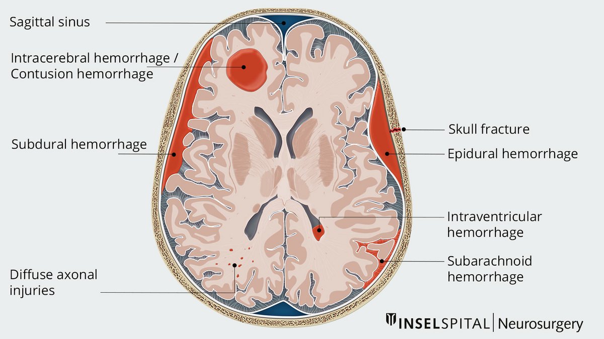
- Epidural hemorrhage (between the skull bones and the dura mater)
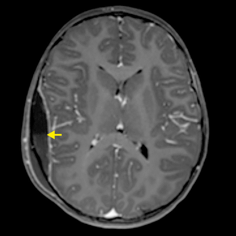
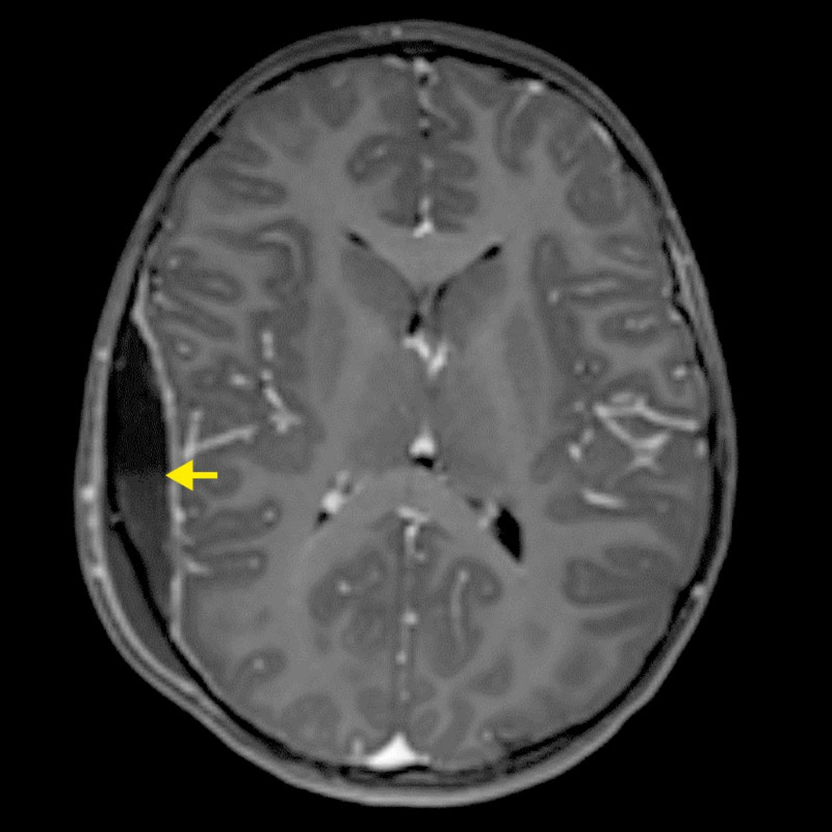
Epidural hematoma. The MRI cross-sectional image with contrast agent shows a bone fracture and epidural hemorrhage following a fall. Bild: Universitätsklinik für Neurochirurgie, Inselspital Bern 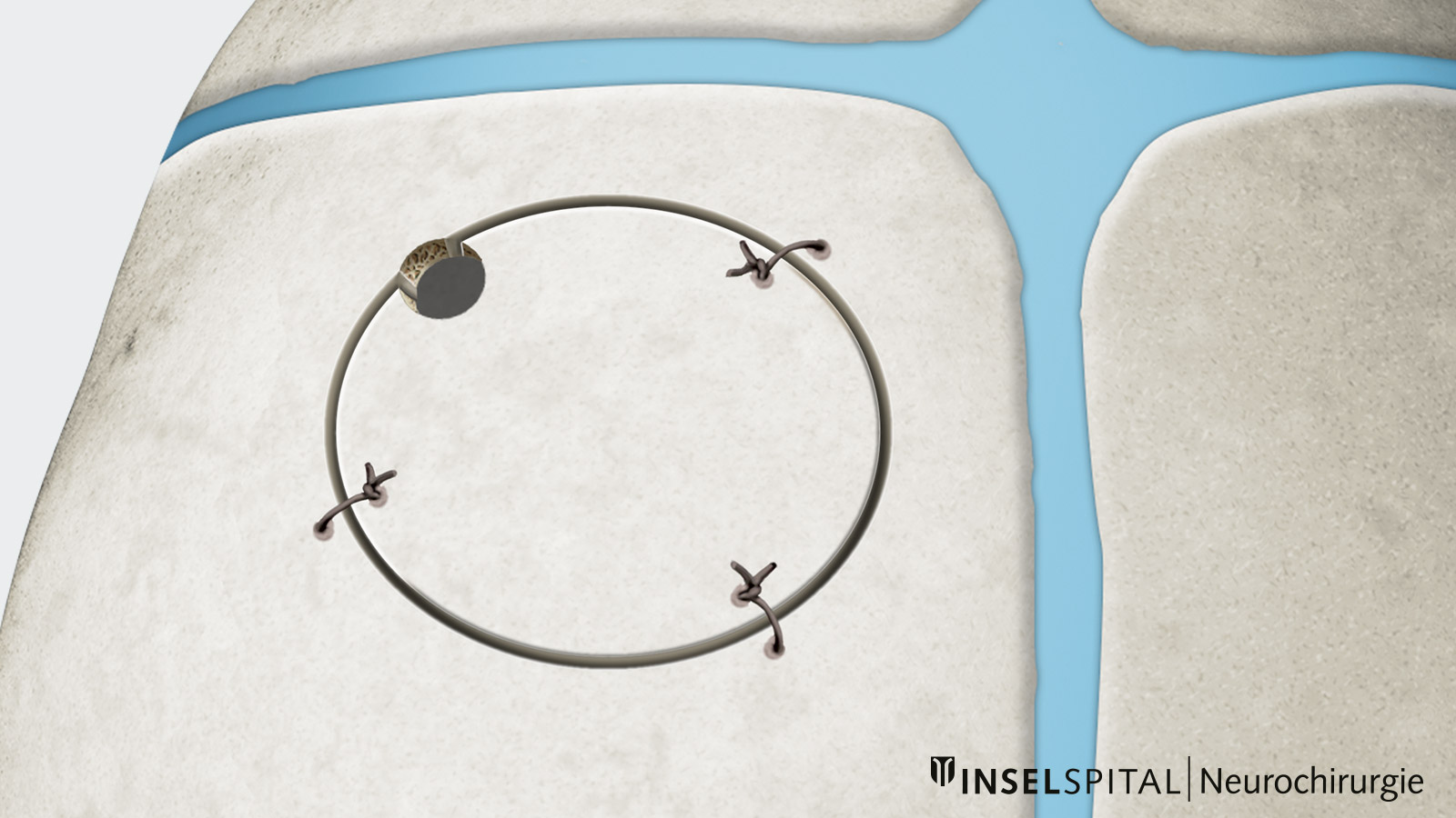
Craniotomy. Drawing of a bone flap that has been repositioned and secured. Bild: Universitätsklinik für Neurochirurgie, Inselspital Bern © CC BY-NC 4.0 An epidural hemorrhage, also known as an epidural hematoma, is a trauma-related bleeding in children and adolescents that relatively often requires surgery. It is usually caused by injury to a meningeal artery beneath a skull fracture.
Nevertheless, a significant proportion of epidural hematomas in children can be treated without surgery and with close follow-up monitoring. Surgery is primarily necessary if the hemorrhage compresses the brain, thereby causing neurological deficits, or if it rapidly increases in size *.
Important: Especially in young children, a significant epidural hematoma may cause virtually no symptoms for an extended period. The absence of symptoms therefore does not rule out such a hemorrhage *.
During surgery, the surgeon opens the skull bone over the site of the hemorrhage, removes the blood clot, and closes the bleeding vessel – usually a small meningeal artery. The removed bone flap is then replaced and secured.
Since the hemorrhage is located outside the brain itself and the brain is usually not damaged in the process, children generally recover very well and completely following timely surgery.
- Subdural hemorrhage (between the dura mater and the pia mater)
A subdural hemorrhage, also known as a subdural hematoma, is usually caused by the rupture of so-called bridging veins. It occurs more frequently in conjunction with a severe, diffuse brain injury than an epidural hemorrhage. Unlike in older patients, subdural hematomas are less common than epidural hematomas in children and adolescents.
If the hemorrhage significantly compresses the brain, surgery is necessary. The procedure is usually performed through a relatively small opening in the skull through which the hematoma is removed.
In cases of extensive bleeding, particularly when contusion hemorrhages are also present, it may be necessary to temporarily remove half of the skull vault. This procedure is called a decompressive hemicraniectomy. It creates the necessary space for the injured and swelling brain.
Once the brain has recovered, the skull vault or a synthetic bone implant is reinserted during a second operation.
- Subarachnoid hemorrhage (in the fluid-filled space surrounding the brain)
- Intracerebral hemorrhage/contusion hemorrhage
As with spontaneous cerebral hemorrhages that occur without prior traumatic brain injury, the size and location of the intracerebral hemorrhage, as well as the patient’s symptoms, determine whether surgery is necessary or whether non-surgical treatment is sufficient.
These hemorrhages are often accompanied by serious brain swelling. The swelling may not reach its peak until several days after the accident. For this reason, prolonged monitoring in the pediatric intensive care unit is sometimes necessary.
- Intraventricular hemorrhage (in the brain ventricles filled with cerebrospinal fluid)
- Diffuse axonal injury (shearing injury)
When the head undergoes rapid acceleration or rotation—such as in a high-velocity or shaking trauma—fine nerve fibers deep within the brain can become overstretched or twisted and tear. This injury is known as diffuse axonal injury (DAI).
Unlike in a classic cerebral hemorrhage, this often results in no significant accumulation of blood. Instead, there may be numerous very small injuries and hemorrhages that are not always visible on a computed tomography (CT) scan. A magnetic resonance imaging (MRI) scan may therefore be necessary to more accurately assess the extent of the injury.
Infants are particularly at risk because their heads are heavy relative to their bodies and their neck muscles are still weak. Diffuse axonal injuries therefore frequently occur as a result of shaken baby syndrome.
Since a diffuse axonal injury (DAI) can lead to long-term cognitive, emotional, or behavioral problems, an early child neuropsychological evaluation (explanatory video in German) and tailored pediatric neurorehabilitation are important. *

When is surgery necessary?
Surgery may be particularly necessary if:
- brain tissue is being displaced
- bleeding is rapidly increasing
- the child’s level of consciousness is deteriorating
- there is a risk of brain tissue becoming pinched
The treatment team makes a case-by-case determination as to whether surgery is required or if intensive care measures are sufficient, and which of these are needed. The team includes pediatric neurosurgery, pediatric intensive care, neuropediatrics, and pediatric surgery, as well as other specialties, depending on the course of the illness and its cause.
Treatment of increased intracranial pressure – intensive care measures
In cases of traumatic brain injury or spontaneous intracerebral hemorrhage, the brain may swell. If this causes the pressure inside the skull to rise too high, there is a risk of further brain damage.
In unconscious or deeply sedated children, increased intracranial pressure cannot be reliably detected externally. For this reason, it is often measured directly using an intracranial pressure (ICP) catheter or an external ventricular drain (EVD). Both are inserted during a brief procedure and allow treatment to be continuously adjusted based on the patient’s progress.
Treatment is carried out in stages – from basic to more intensive measures. As soon as the child’s condition stabilizes, the measures are gradually scaled back *.
Basic measures
The upper body is slightly elevated and the head is positioned upright. Fever is treated. In addition, the treatment team ensures stable circulation and a balanced fluid and electrolyte status.
Pharmacological reduction of intracranial pressure
Certain infusion solutions temporarily remove fluid from the swollen brain tissue, thereby reducing intracranial pressure.
External ventricular drainage (EVD)
A thin catheter is inserted into the brain ventricles, which are filled with cerebrospinal fluid (CSF). This allows excess cerebrospinal fluid to be drained, thereby reducing pressure in the head. At the same time, the drainage system enables continuous monitoring of intracranial pressure.
Induced coma
If these measures are insufficient, the child can be placed in a deep, controlled state of sleep. This reduces the brain’s energy requirements. The treatment is carried out under close monitoring in the pediatric intensive care unit and is scaled back as soon as the situation stabilizes. Brain activity can be monitored using electroencephalography (EEG) – continuously if necessary.
Decompressive craniectomy
As a last resort, part of the skull bone may be temporarily removed. This provides more space for the swollen brain.
The removed bone is stored under sterile conditions and is usually reinserted several months later, once the swelling has subsided. If the patient’s own bone is no longer suitable for this purpose, a custom-made artificial implant can be used instead. Since the benefits and risks of this major surgery vary from person to person, the procedure is carefully evaluated on a case-by-case basis *.
What is different in children compared to adults?
The treatment of traumatic brain injury must be tailored to the child’s age and developmental stage. This applies to diagnosis, monitoring, treatment, and follow-up care. Some important differences are:
- Symptoms and Treatment: The symptoms and severity of a brain hemorrhage may differ from those in adults. Therefore, the reasons for surgery and the surgical technique may also differ.
- More flexible skull bones: Especially in infants and toddlers, the skull bones are still softer. This can lead to characteristic indentations, known as ping-pong fractures. There are also differences in the healing process. For example, in young children, a growing fracture must be ruled out as the condition progresses.
- Lower blood volume: Children have less blood in their bodies overall. Especially in toddlers, even a small amount of blood loss can therefore quickly become significant.
- Age-appropriate surgical techniques: In adults and adolescents, a repositioned bone flap is usually fixed with titanium plates. In younger children, depending on their age, sutures, bioresorbable (self-dissolving) implants («sugar plates»), or other techniques that account for continued growth may also be used. In some cases, different materials may be used for bone replacement than those used in adults.
- Recovery and follow-up care: The child’s brain possesses a high degree of adaptability, known as plasticity. For this reason, pediatric neurorehabilitation, pediatric neuropsychology, and interdisciplinary care are particularly important.
What is the prognosis after a traumatic brain injury?
What is the prognosis after a traumatic brain injury?
Most children with a mild traumatic brain injury are completely symptom-free again after a few weeks to a few months.
Even after more severe injuries, children can recover well thanks to the high adaptability of the child’s brain. However, the prognosis depends on the severity of the brain injury, the type and extent of any bleeding, and any accompanying injuries.
Pediatric neurorehabilitationand pediatric neuropsychology play an important role in long-term recovery.
Why you should have your child treated at Inselspital
Children with traumatic brain injury receive round-the-clock care at Inselspital from an experienced interdisciplinary team. Depending on the situation, the following departments are involved:
- Pediatric Neurosurgery
- Pediatric Emergency Medicine
- Pediatric Critical Care Medicine
- Neuropediatrics
- Neuroradiology
- Pediatric Surgery
- Pediatric Neuropsychology
- Pediatric Neurorehabilitation
All treatment steps are planned individually by the interdisciplinary team in collaboration with the parents.
As a university medical center, we have the experience and infrastructure to treat even complex and rare injuries, such as the most severe traumatic brain injuries, in children of all ages—from premature infants to adolescents. Thanks to close collaboration among various specialties, we can also provide comprehensive care for severe concomitant injuries.
Diagnostics, surgical techniques, implants, and follow-up care are tailored to the child’s age and specific anatomy.
Our in-house pediatric neurorehabilitation program is also a major advantage for successful treatment.
-
Lumba-Brown A, Yeates KO, Sarmiento K, et al. Centers for Disease Control and Prevention Guideline on the Diagnosis and Management of Mild Traumatic Brain Injury Among Children. JAMA Pediatr. 2018 Nov 1;172(11):e182853. doi: 10.1001/jamapediatrics.2018.2853. Epub 2018 Nov 5. Erratum in: JAMA Pediatr. 2018 Nov 1;172(11):1104. doi: 10.1001/jamapediatrics.2018.3868.
-
Roche S, Crombé A, Benhamed A, Hak JF, Dabadie A, Fauconnier-Fatus C, Rega A, Pech-Gourg G, Tazarourte K, Seux M, Acquier A, Gorincour G. Risk Factors Associated with Traumatic Brain Injury and Implementation of Guidelines for Requesting Computed Tomography After Head Trauma Among Children in France. JAMA Netw Open. 2023 May 1;6(5):e2311092. doi: 10.1001/jamanetworkopen.2023.11092.
-
Lumba-Brown A, Yeates KO, Sarmiento K, et al. Diagnosis and Management of Mild Traumatic Brain Injury in Children: A Systematic Review. JAMA Pediatr. 2018 Nov 1;172(11):e182847. doi: 10.1001/jamapediatrics.2018.2847. Epub 2018 Nov 5. Erratum in: JAMA Pediatr. 2018 Nov 1;172(11):1104. doi: 10.1001/jamapediatrics.2018.3879.
-
Leddy JJ. Sport-Related Concussion. N Engl J Med. 2025 Jan 30;392(5):483-493. doi: 10.1056/NEJMcp2400691.
-
Wang H, Zhou Y, Liu J, Ou L, Han J, Xiang L. Traumatic skull fractures in children and adolescents: A retrospective observational study. Injury. 2018 Feb;49(2):219-225. doi: 10.1016/j.injury.2017.11.039. Epub 2017 Nov 29. Erratum in: Injury. 2018 Jul;49(7):1365. doi: 10.1016/j.injury.2018.05.009.
-
Skadorwa T, Zygańska E, Eibl M, Ciszek B. Distinct strategies in the treatment of epidural hematoma in children: clinical considerations. Pediatr Neurosurg. 2013;49(3):166-71. doi: 10.1159/000359954. Epub 2014 Apr 8.
-
Greenberg JK, Stoev IT, Park TS, Smyth MD, Leonard JR, Leonard JC, Pineda JA, Limbrick DD. Management of children with mild traumatic brain injury and intracranial hemorrhage. J Trauma Acute Care Surg. 2014 Apr;76(4):1089-95. doi: 10.1097/TA.0000000000000155.
-
Kochanek PM, Tasker RC, Bell MJ, Adelson PD, Carney N, Vavilala MS, Selden NR, Bratton SL, Grant GA, Kissoon N, Reuter-Rice KE, Wainwright MS. Management of Pediatric Severe Traumatic Brain Injury: 2019 Consensus and Guidelines-Based Algorithm for First and Second Tier Therapies. Pediatr Crit Care Med. 2019 Mar;20(3):269-279. doi: 10.1097/PCC.0000000000001737.
-
Di Cosmo L, El Choueiri J, Pellicanò F, Ozsut O, Antonino JR, Feffer EB, Ghosh S, Darwiche A, Imbrogno CP, Cuervo SN, Chibbaro S, Zaed I. Surgical Versus Medical Management for Severe Pediatric Traumatic Brain Injury: A Systematic Review and Meta-Analysis. Neurosurgery. 2026 Mar 10. doi: 10.1227/neu.0000000000003974. Epub ahead of print.